The Inside Story: Computerised Tomographic Examination of a Middle Bronze Age Cremation Urn From Barton-Under-Needwood, Staffordshire.
By Duncan J. Robertson
Introduction
For possibly the first time, a Middle Bronze Age cremation urn and its undisturbed contents have been examined by CT scan. The procedure was part of a MAP 2 assessment exercise, performed at the University of Bradford in conjunction with Airedale General Hospital, following the urn's recovery during a rescue excavation. The use of Computer-aided Tomography in archaeology is not new; however, it has mostly been confined to the study of Egyptian sarcophagi, wrapped and embalmed mummies, biological anthropology, biomechanics, the diagnosis of palaeopathological conditions and, most recently, the examination of the prehistoric frozen corpse discovered on the Tyrolean Alps, the so-called "�tzi". Anderson and Fell's pioneering article (1995) promoted the potential of the application of this technique to a different type of closed funerary context, that of the cremation urn and suggested that the technique is a useful tool for rapidly assessing the composition of an urn's fill and the precise nature of its contents. However, this represents only a fraction of the potential of CT scanning technology for the recording, examination and storage of cremations from archaeological sites.
Background
Rescue excavation by archaeological contractors, Gifford and Partners, at the site of Barton-Under-Needwood, Staffordshire (NGR 2084 1880), prior to the development of an industrial estate in the summer of 1996, revealed a flat cremation cemetery in the vicinity of an earlier ring cairn. Some of the cremations were deposited in urns and the remainder were in non-urn contexts. A group of three Middle Bronze Age urns of Deverel Rimbury type were selected for laboratory micro-excavation (fig.1).
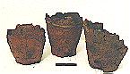 Figure 1
The three excavated urns following consolidation, microexcavation and conservation at the department of archaeological science, University of Bradford. Urn number 742 is to the right of the photograph. The scale is 10cm. (Photograph courtesy of Yannick Minvielle-Debat) Click for full size image.
Figure 1
The three excavated urns following consolidation, microexcavation and conservation at the department of archaeological science, University of Bradford. Urn number 742 is to the right of the photograph. The scale is 10cm. (Photograph courtesy of Yannick Minvielle-Debat) Click for full size image.
This procedure formed part of an assessment exercise to determine the potential of the cremated remains for further specialist osteological study and to allow the urns to be conserved and reconstructed. Following initial consolidation, one of this group of urns (number 742) was selected for CT scanning in order to examine its contents and structure prior to micro-excavation.
In Anderson and Fell's study (1995), intact Roman vessels were analysed. In this instance, the urn was prehistoric and incomplete, hence the need for consolidation and careful packing before transport to the hospital where it was to be scanned. A further and more fundamental contrast between their work and the present study was that this was not simply an osteoarchaeological exercise, but one to assess the potential of CT scanning for the purposes of the project conservator. The results of the scan allowed the assessment of the precise condition of the urn so that it could be efficiently reconstructed. The presence of any material within the fill that required the attention of the conservator could also be identified prior to micro-excavation and the necessary procedures set up at an early stage. The justification for the scan, therefore, was that it could assist in the recording, interpretation, excavation, conservation and reconstruction of the urn and its contents not just for osteological purposes, but as an additional tool for the conservator.
Method
Following consolidation, the urn was initially taken for X-radiography at the School of Health, University of Bradford. The resulting image superimposed the structures apparent in the urn into two dimensions. Ultimately, this plate was of limited use in the interpretation of the contents of the urn as it proved impossible to identify the nature or three-dimensional location of any of the structures present with any degree of accuracy.
The urn was transferred to the CT Scanning department at Airedale General Hospital, where it was placed in the chamber of a Siemens Somatom HiQ-S scanner normally used for scanning sections of the internal organs of live patients. The urn was treated as a head scan and images were taken as a series of vertical slices at 10mm intervals at 133KV and 175mA as the artefact passed through the scanner. This produced a total of 29 scanned images which were subsequently reproduced by a Siemens laser imager onto Dupont Chronex Safety film. The immediate advantage over the X-ray was that the images were initially displayed on a computer screen and could be manipulated to show the preferred image, which was then printed out. Depending on the scan type, three-dimensional images or slices constructed perpendicular to the original axis of the scan could be reproduced.
Results
The plates produced in this instance illustrated the composition of the urn's fill in a sequence of slices. There was no superimposition as experienced in the X-ray and the contrast of the image was also superior. The condition of the urn was clearly illustrated, with the walls and base observed to be fractured throughout, requiring considerable attention during micro-excavation and the subsequent reconstruction (fig. 2).

 Figure 2 (left)
Initial scan of urn 742, illustrating the degree of fragmentation of the sides of the vessel. Figure 3 (right) Central scan image of the urn and its contents, illustrating the stratigraphic distribution and fill types. Note the darker areas of radiolucency in the top right of the image, which correspond to voids created by earthworm activity. Click for full size images.
Figure 2 (left)
Initial scan of urn 742, illustrating the degree of fragmentation of the sides of the vessel. Figure 3 (right) Central scan image of the urn and its contents, illustrating the stratigraphic distribution and fill types. Note the darker areas of radiolucency in the top right of the image, which correspond to voids created by earthworm activity. Click for full size images.
Three layers within the urn were clearly identified on the plate and were found to consist of the lowermost matrix, containing the cremated bone, a central gravel-based material and the upper layer of stone-free silt (fig. 3).
The clarity obtained was demonstrated by the appearance of voids in the upper layer that corresponded to worm infiltration. The scan therefore appeared to have the potential to highlight post-excavation disturbance by soil fauna and this was demonstrated when the contents of the urn were excavated.
Skeletal elements were only identifiable as fragments of long bone shaft (fig. 3); however, the prior identification of the extent of this layer of bone proved of value when it came to emptying the contents of the urn. The osteologist was not working blind and did not have to produce a section drawing of the contents, as 29 highly accurate images already existed, complete with scale. The speed at which the urn could be emptied was increased with the aid of this additional data.
Discussion
Identification of cremated skeletal elements in the CT scan images produced in this assessment was limited to examples of long bone shafts caught in section, cranial vault fragments and vertebral bodies. This was due to the two-dimensional nature of the slices produced. However, the potential exists for the three-dimensional reconstruction of the contents of an urn as demonstrated by Baldock et. al. (1994). In this study, an unopened Egyptian sarcophagus was CT scanned and then, with the aid of powerful computers, a three-dimensional image was constructed. The mummified corpse was virtually unwrapped without damaging physical intervention. Following this 'unwrapping', the soft tissues and skeletal elements were stripped away to reveal the dentition. The resolution of this final image proved sufficient for an oral biologist to age the individual using the stage of apical closure of the molar tooth roots.
This degree of resolution can theoretically be applied to a cremation urn in order to 'excavate' the contents without actually disturbing them. In an ideal world, a three-dimensional manipulation of the image on a computer screen should enable the osteologist to identify the skeletal elements present and use the results to reconstruct biological details of the individual(s) contained within the urn and the order in which the remains were deposited.
CT scanning is a non-destructive means by which one can accurately pinpoint the location of the urn's contents in three dimensions. Points of weakness in the urn can be identified by the conservator as requiring extra attention and the contents of the urn can be assessed, allowing the presence of artefacts within the fill to be identified and, if necessary, excavated accordingly. CT scans can positively identify the presence of stone, bone, metal, soil layers, voids and differences in compaction, as well as the degree of fragmentation of both bone and the vessel itself. The results can indicate the best strategy for micro-excavation and who should perform this task: either a finds assistant, in cases where the urn is shown to be empty; a conservator, in examples where the urn or its contents require the attention of such specialist skills; or, finally, an osteologist, who can perform the final analysis of the urn's contents in order to acquaint him- or herself with the skeletal remains and the condition in which they were recovered. For cremation urns, CT scanning is superior to Magnetic Resonance Imaging (MRI) as ferrous metals present in the fill of an urn can be ripped out of the matrix by the force of the magnetic field in this type of scanner, thus destroying the integrity of the urn and potentially damaging an expensive item of equipment (S. Milner pers. comm.).
With the increasing need for specialists to provide post-excavation assessment reports, CT scanning produces fast results. Urns need not be excavated if they prove empty and a strategy can be formulated in advance to allow the most archaeologically informative sample of urns to be more fully analysed within the time and budget constraints of a post-excavation project. In this example, the unscanned urns took substantially longer to excavate than the scanned urn, as the osteologist was effectively working blind. This technology is accessible to the archaeologist through contact with hospital scanning departments, although the work has to be carried out without interfering with patient schedules and this can lead to scanning at unsociable times of day. However, this is a small price to pay in order to ensure that studies such as this one do not remain solitary attempts to realise the potential of CT scanning technology.
Conclusions
CT scanning technology currently available to archaeologists remains a useful time and labour saving device, although its application to rescue archaeology can be costly, particularly for underfunded units or under-budgeted projects. However, its use can lead to a more streamlined project and the efficient use of specialist time and labour, which ultimately equals a cost-effective post-excavation operation. In terms of conserving our archaeological heritage and allowing the remains of the dead to lie undisturbed, the use of the CT scanner potentially allows a compromise to be established between the needs of a scientific discipline and the respect that must be shown to the deceased; although three-dimensional reconstruction of the contents of cremation urns may yet be many years away. However, for archaeology to be seriously regarded as a scientific discipline, the potential of new technologies, such as CT scanning, to the study of a wide range of materials should be explored and applied to both research and rescue projects.
In terms of research, the application of powerful computers to the construction and manipulation of precise three-dimensional images will breathe new life into the recording, examination, presentation and storage of the class of artefacts under discussion here, and will ultimately negate the need to actually excavate their contents. Reburial of the urns could become a realistic alternative to long term storage in museum collections, with all the data from the CT scan being stored in a database and accessible as either the raw data for the researcher, or as an educational three-dimensional reconstruction for the public. There would be no need for the public display of cremated human remains, which some consider unethical. The storage of virtual as opposed to real artefacts may also reduce the pressure on national and regional museums, whose curatorial facilities are currently stressed to the limit, by replacing a bulky urn with a digital alternative which takes up a fraction of the space. The use of CT scanning and modern computing technology therefore can potentially be of benefit to archaeology in the long term. This potential just needs realising.
Acknowledgements
I would like to thank those involved with the assessment of the Barton-Under-Needwood project: Mr A. Martin of Gifford and Partners; Mrs. A. Boylston, Calvin Wells Laboratory, Bradford University Department of Archaeological Sciences.
To Mr. S. Milner, Head of Division of Radiography, Bradford School of Health, University of Bradford and Ms. A. M. Duchesne, Superintendent III radiographer, Airedale N.H.S. Trust Hospital, for the opportunity to use their expertise and X-ray and CT scanning facilities.
Finally to Dr. Martin Evison, for his comments on an earlier draft of this paper and to Yannick Minvielle-Debat, Conservator at the University of Bradford Department of Archaeological Sciences, for comments on earlier drafts of this paper, consolidating the urn, organising the CT scan and transporting the urn and this osteologist to the necessary institutions.
References
Anderson, T. and Fell, C. 1995. Analysis of Roman Cremation Vessels by Computerised Tomography. Journal of Archaeological Science 22:609-617
Baldock, C., Hughs, S., Whitaker, D., Davies, R., Taylor, J., Spencer, A. J., Sofat, A. and Tonge, K. 1994. 3-D Reconstruction of Ancient Egyptian Mummy using X-ray Computer Tomography. Scope 3 (2) 21-24
About the author: Duncan Robertson has recently completed the M.Sc. in Osteology, Palaeopathology and Funerary Archaeology, run jointly by the Universities of Sheffield and Bradford, and is currently working for
ARCUS as a contract osteoarchaeologist. He is a connoisseur of fine ale, Formula 1 and his favourite Spice Girl is Geri.
�Duncan Robertson 1997
� assemblage 1997