
Estimating Osteological Health in Ancient Egyptian Bone via Applications of Modern Radiological Technology.
Estimating Osteological Health in Ancient Egyptian Bone via Applications of Modern Radiological Technology.
This paper offers a process evaluation of the use of dual energy x-ray absorptiometry (DXA) in the study of ancient human remains. The study was undertaken to assess the potential use of the DXA technique as a non-invasive and non-destructive method of assessing bone health in an ancient population: poor diet, for example, could reasonably be expected to affect bone density.
Background
For the forensic archaeologist, osteological remains form the most important source of primary paleopathological evidence. Samples of bone 'transcend in abundance any other kind of evidence' (Wells, 1964). It must be acknowledged that bones do not endure the millennia unchanged, many factors contribute to the eventual destruction, but there remains much that osseous material can reveal about the individual and the biocultural context in which they lived (Mays, 1998). There is evidence that osteoporosis (bone thinning, which is idiopathic or secondary to other disease) and osteopenia (decrease in bone mass below the norm) were present in ancient Egyptian populations (Dequeker et. al. 1997). For these disorders to be apparent on simple radiological examination there must be significant loss, i.e. 40% or more, of bone density, which makes this technique generally unsuitable for archaeological materials. Whilst work has been carried out on ancient Egyptian remains to assess osteopenia via stable isotope analysis (White & Armelagos, 1997), the technique requires destruction of a sample of bone. Likewise the assays of alkaline phosphatatse (an enzyme that is raised whenever there is bone destruction present) requires destruction of the organic material. This means that at present there is no widespread non-destructive method of achieving a clear and accurate understanding of the epidemiology of osteoporosis and osteopenia in ancient populations. However, a radiographic technique that potentially allows for assessment of bone mineral content and bone density in a non-destructive manner is dual X-ray absorptiometry (DXA), which is utilised in modern radiology to assess bone density as well as to facilitate the diagnosis and risk of osteoporosis.
The role of the DXA technique in the field of paleoradiology can be argued to have less focus upon diagnosis and more upon straightforward investigation. The procedure had not, prior to this process evaluation study, been used upon ancient Egyptian material and indeed is not a widely used technique within paleopathology. Lees et al. (1993), in their seminal paper, utilised DXA in a comparison of the Bone Mineral Density (BMD) of modern day women with 18th and 19th century skeletal remains from the crypt of Christ Church, Spitalfields. They found significant bone density in their sample of 18th and 19th century women, whom they postulated had been predominantly factory workers. In many cases the bone density of the ancient samples was better than the modern population and this was thought to be a result of better diet in regards to the Christ Church sample (fresh vegetables, less processed food etc.) (Lees et. al. 1993).
Sample
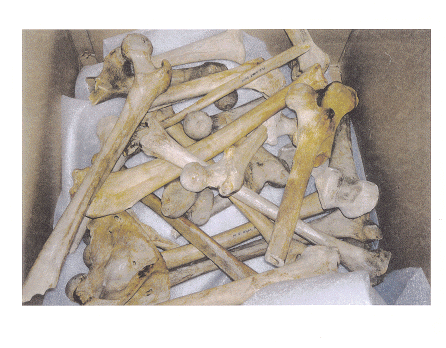
The material utilised within this study is considerably older than that from Christ Church, Spitalfields, having been drawn from the Elliot Smith Egyptian collection of the Manchester Museum (Figure 1). The source of the osteological material used in this study was from burials at fifty-seven sites ranging from pre-dynastic to Roman times, a time span that covers the whole of ancient Egyptian history from 5500 BCE to 395 CE. (Nunn, 1996) This would mean that the bones used for this study could be anything from 2000 to 7000 years old.
Figure 1: The Sample Pool
As Figure 1 shows, the sample pool contained femora in various states of anatomical completeness. Femora were used in preference to other bones since anatomical markers, such as femoral neck angle, can provide insights into the activity of the individual (Spencer Larsen, 1997). It became clear that as long as there was a reasonable amount, i.e. 10-15 cm or more, of femoral shaft in existence, the sample was appropriate for the purpose of scanning. From the sample pool available, three femora were picked at random. The bones were given names since they were entered into the DXA computer as 'real' patients. The second specimen was given two names because it was scanned twice with different genders entered into the computer. All specimens scanned were anatomically complete from head of femur to femoral condyles. The osteological samples that were utilised in this study were examined macroscopically and their dimensions recorded. The description and morbid measurements of the bones sampled are outlined in Table 1. This dimension mapping covered areas of importance on the sample (see for anatomy of femur).
|
E. Femora |
Amelia/Hapi |
Maat |
|
|
Classification |
Left Femur |
Right Femur |
Left Femur |
|
Length |
402 mm |
406 mm |
401 mm |
|
Circumference of femoral shaft |
90 mm |
80 mm |
725 mm |
|
Circumference of femoral head |
105 mm |
102 mm |
95 mm |
|
Diameter of femoral head |
44 mm |
43 mm |
37 mm |
|
Length of femoral neck: Greater trochanter |
20 mm |
10 mm |
5 mm |
|
Length of femoral neck: Lesser trochanter |
30 mm |
15 mm |
15 mm |
|
Angle of femoral neck |
125º |
140º |
120º |
|
Width across trochanters |
60 mm |
80 mm |
70 mm |
Table 1: Morbid measurement of osteological specimens
Three of the measurements, femoral head diameter, femoral neck angle and femoral shaft length were used to extrapolate gender, activity levels and height (Spencer Larsen, 1997; Genovés, 1969; Trotter et. al. 1952). The measurements of the femoral head suggested that all of the specimens used were female (Chamberlain, 1994). There is evidence to suggest that femoral head size has remained more or less the same in relation to body mass throughout the last 1500 years of human history (Spencer Larsen, 1997). Changes in loading of the body can manifest themselves as alterations in trabecular bone at the articular joints. Differences in the angle of the femoral neck are less individualised and reflect more the development of a society with foragers having the lowest femoral neck shaft angles and urban dweller having amongst the highest, this reflects the amount of activity and muscle forces used (Spencer Larsen, 1997). The bone with the poorer density readings (Amelia/Hapi) did have a femoral neck angle that suggested a reasonable sedentary lifestyle, but without accurate biographical data it is difficult to assess the validity of this measurement.
Methodology

The samples were scanned using a DR-2000 Plus High Resolution Bone Densitometer (Figure 2). This machine uses a low radiation source and an in-built computer to assess both bone mineral content (BMC) and bone mineral density (BMD). Each scan was completed in less than two minutes.
Figure 2: The DR-2000 Plus High Resolution Bone Densitometer
One of the difficulties surrounding the issue of estimating bone mineral density via a DXA scan on live patients is the non-uniformity of soft tissue. In this study, the reverse applied and difficulties were anticipated because there was no soft tissue present. Given that air is radiolucent and that the scanner was calibrated to accommodate soft tissue this absence of would have severely compromised the results. On average, the thickness of soft tissue overlying bone is approximately 15cm.Lees et. al. (1993) immersed their samples in 15 cm of water. I was reluctant to do this, primarily because of the age of the osteological material being used. Also, the damage that had been noted on the outer cortex of the bone, which had exposed the underlying trabecular bone, would have allowed the water to seep into the bone matrix potentially damaging the specimen. The radiographer suggested that bagged rice, which has been used to good effect with live patients whose extreme thinness may have compromised their scan result, would serve as an adequate substitute. The dry bone was supported on eight one kilogram bags of rice and two more bags covered the femoral head and neck (Figure 3). Measurement of this set-up indicated that the requisite fifteen cm. soft tissue equivalent had been achieved.
 Figure 3: Soft Tissue Simulation
Since it has been contended that only 10% of the ancient Egyptian population survived beyond the age of fifty (Worth Estes, 1993) each bone was scanned three times, as a thirty year old, a forty-five year old and a fifty year old. The data of interest is expressed as 'T' and 'Z' scores. The T score compares the bone mineral content of the specimen with the peak bone mass of a healthy thirty year old. The 'Z' score compares the BMD with the norm for the stated age. Both results are expressed as percentages.
Results
The results that we obtained from the three samples are outlined below. (Table 2)
|
Scan 1: 50 Years Old |
Scan 1: 45 Years Old |
Scan 1: 30 Years Old |
||||
|
|
BMC |
BMD |
BMC |
BMD |
BMC |
BMD |
|
|
T Score |
Z Score |
T Score |
Z Score |
T Score |
Z Score |
|
E. Femora |
100% |
112% |
100% |
109% |
100% |
100% |
|
Amelia |
93% |
105% |
94% |
102% |
94% |
95% |
|
Hapi |
85% |
98% |
85% |
95% |
85% |
89% |
|
Maat |
88% |
100% |
87% |
95% |
86% |
87% |
Table 2: DXA measurements for Bone Mineral Content (BMC) & Bone Mineral Density (BMD).
Taphonomic Problems
Osteological material can survive the passage of time better than almost any other biological material, however, the integrity of a bone sample can be compromised by soil acidity, water damage, temperature and soil dwelling micro-organisms (Mays, 1998). Trabecular bone appears to decay faster than cortical bone in inhospitable soils, the compact bone of the shaft of long bones will survive whilst the trabecular bone of the condyles or head will not (Mays, 1998). Acidic soils and/or the acidic microenvironment, caused by micro-organisms living in the soil, contribute to either the degradation of hydroxyapatite (the main constituent of bone mineral) or its conversion to brushite. Brushite, another mineral, occupies more space than hydroxyapatite and tends to crack and splinter the bone. Water provides a medium for the transport of ions to and from bone and contributes in a major way to the chemical degradation of osteological materials. Higher temperatures facilitate all of these processes and also contribute to the destruction of collagen, which in turn can start to dissolve bone minerals (Price, 1989).In addition, post-mortem damage, either at the time of excavation or from subsequent poor storage and conservation, can lead to loss of bone. In certain instances this will either, affect the quantitative data obtained, as happened in the case of the 'Maat' specimen, or render the specimen unsuitable for density measurement.
However, it can be argued that of all osteological material, ancient Egyptian bones may be amongst the best preserved in terms of their mineral content. Guarded from soil acidity by their rock cut tombs, encased within their protective sarcophagi and benefiting from the clement environment and the religion and science of preservation of the body, the ancient Egyptians ensured skeletal survival. Whilst the preservation of bone integrity could have been identified as a confounding variable within this study, the care that the ancient Egyptians lavished upon their dead meant that the specimens used were virtually osteologically intact.
Process evaluation
Two important factors were highlighted as potential threats to validity and reliability when carrying out multiple scans on one specimen. Occasionally the scanning arm of the DXA equipment did not reposition itself exactly in the spot from which the first scan was undertaken. The differences in readings were minimal, however, and usually this is acknowledged as a confounding variable that can be discounted. However, there was demonstration of machine variability when scanning the 'Maat' specimen.
Figure 4: Ward's Triangle

On scanning the 'Maat' bone for the third time, the machine moved the Ward's triangle region (see Figure 4) from the base of the femoral head, where it had been in the two previous scans, to slightly above the intertrochanteric region. This would clearly have affected the quantitative data, on rescan however, the Ward's triangle region was comparable with previous scans and we concluded that the anomalous scan had been a system glitch. This problem emphasised the importance of checking and rechecking the computer data between scans.
The radiographer who was associated with this study highlighted the second threat to validity, that of operator variability. She pointed out that different results could be obtained from the same patient on the same day if two different radiographers carried out the scans. This can usually be explained away by the differences in identifying the 'area of interest' on the monitor screen. This variability is not the same as error in that the differences are often minute, but they are present. In a situation such as the one outlined for this work, this may be an issue when the same sample is being scanned more than once with different age or gender parameters. It is, therefore, recommended that the same radiographer carry out all of the DXA scans. The process is also facilitated if that radiographer is skilled in the use of this equipment. This will help to ensure continuity of identification of area of interest and will, in turn, increase the reliability of the reading obtained.
Given that the work carried out by Lees et al. (1993) is the only other paleoradiological application of DXA technology, the variations between the two techniques must be highlighted. Wahner and Fogelman (1994) noted that the Hologic instruments (as used in this study) showed a 29% lower calculation of the bone mineral density values of Ward's triangle when compared with a Lunar DPX (as used by Lees et. al. 1993). In addition, Wahner and Fogelman state that the Hologic equipment measures bone mineral content as lower than the Lunar system. It was also noted that, with the exception of the Ward's triangle, the region of interest outlined by the Lunar DPX-L was smaller. These comparisons, by focusing upon smaller areas and recognising smaller changes in mineral content and density, would tend to indicate a greater degree of precision in the Hologic equipment. Whilst these differences may be insignificant, it is recommended that any DXA examination is carried out on one machine only to address this variability.
Conclusion
This study has shown that dry bone scanning can provide relevant and quantifiable data. Unlike Lees et al. (1993), who were able to immerse their osteological specimens in water in order to replicate soft tissue thickness, this study has shown that bagged rice is an acceptable alternative for this necessary simulation. This technique has the added advantage of keeping the bone dry, an important consideration in ensuring integrity and survival of the specimen. Lees et al. (1993) noted that the bone density of the Spitalfields material was superior to that of the modern women. This was also indicated by the results achieved by this study.
Testing the DXA process on unprovenanced material could be seen as a weakness of this study. However, to offset these potential problems the computer was given three different ages for each of the bone specimens scanned. On reflection, it would have been more appropriate to keep to a ten year time span, i.e. thirty, forty and fifty years old since a five year time gap does not make a significant difference to the results that are obtained. This study has clearly demonstrated that for the DXA technique to produce results of any meaning there must be some background information regarding the osteological material under investigation. The samples used for this investigation could have lived at any time on a 5000 year long time continuum. It was not known if the specimens were from the same era, the same region, the same social class or even the same gender (although the dimension mapping did allow us to speculate on that issue). This made comparison of results inappropriate. However, if biographical information is available there is an opportunity for DXA to illustrate changes in bone mineral density across genders, ages and social class. A simple indication of gender may also allow the estimation of BMD to be used to approximate the age of the individual when they died. Bone density has already been used, with other techniques such as cranial suture closure, as a tool for estimating age at time of death. Known as the 'complex method' it is widely used in Continental Europe and involves studying the age related loss of trabecular bone either by x-ray, which is less than accurate, or by sectioning the bones, which is destructive (Spencer Larsen, 1997).
The integrity of the sample is important, significant loss of trabecular bone post-mortem will affect the density of the sample. Poor storage technique may cause loss of cortical bone exposing the more delicate trabecular bone, which will subsequently crumble. Whilst bone that is fractured can successfully undergo DXA examination, process evaluation of the DXA technique suggests that a minimum of 10 cm should exist below the lesser trochanter for the measurements to be valid and mineral density comparisons to be made.
Following both outcome and process evaluation of DXA, it can be argued that there is a role for the technique within forensic archaeology as a whole. The process evaluation has demonstrated that assessment of both bone mineral content and total bone density is possible. The quantitative data that is obtained is meaningful and describes the osteological status of the individual. It has been highlighted that supporting biographical data potentially enhances the validity and reliability of the technique. The strength inherent in the DXA technique is that it is non-invasive and non-destructive, both important issue when one is dealing with a finite amount of ancient human material.
Indications for further study
Having established that dry bone DXA investigation can provide an assessment of bone mineral content and total bone density in ancient osteological specimens, there are numerous indications for further study utilising the DXA process. This method is also useful for intra – and interpopulation comparisons, however it must be born in mind that the measurement will always be against a modern population. If the sample under consideration has rich biographical material it may be possible to make inference regarding lifestyle, activity levels and nutritional status and this should be investigated further. There is also evidence that suggests, in Northern European populations at least, that bone mass varies over the course of the year (Vanderschueren et. al. 1991). Subject to further study this could provide further clues as to season of death.
Issues surrounding the DXA examination of wrapped mummies encased within sarcophagi also require further investigation. It is unknown what effect dead air within the sarcophagus may have upon the DXA readings. It would also be interesting to discover whether enclosure within a sarcophagus relieves one of the need to simulate soft tissue with rice bags. The scanner utilised in this study can also carry out full body mineral measurements. This should be subjected to an outcome and process evaluation similar to the one carried out for this study.
References
Chamberlain, A. 1994 Interpreting the Past: Human Remains. London: British Museum Press.
Dequeker, J., Ortner, D.J., Stix, A.I., Cheng, X.Brys, P., Boonen, S. 1997 Hip Fracture and osteoporosis in a XIIth dyasty female skeleton from Lisht, Upper Egypt. Journal of bone and Mineral Research. 12(6):881 – 888.
Genovés, S. 1969 Sex determination in early man. In: Science in Archaeology. A Survey of Progress and Research. Second Edition. London: Thames & Hudson. pp 429 - 440
Lees, B., Molleson, T., Arnett, T.R., Stevenson, J.C. 1993 Differences in proximal femur bone density over two centuries. The Lancet March 13 341: 763 – 675.
Mays, S. 1998 The Archaeology of Human Bones. London: Routledge
Nunn, J. 1996 Ancient Egyptian Medicine. London: British Museum Press.
Price, T.D. 1989 Bones, chemistry and the human past. In: The Chemistry of Prehistoric Human Bone. Cambridge: Cambridge University Press.
Spencer Larsen, C. 1997 Bioarchaeology. Interpreting Behaviour from the Human Skeleton. Cambridge: Cambridge University Press.
Trotter, M., Gleser, G.C. 1952 Estimation of stature from long bones of American Whites and Negroes. American Journal of Physical Anthropology 10: 463 - 514.
Vanderschueren, D., Gevers, G., Dequeker, J., Geusens, P., Nijs, J., Devos, P., De Roo, M., Bouilon, R. 1991 Seasonal variation in bone metabolism in young healthy subjects. Calcified Tissue International. 49: 84 – 89.
Wahner, H.W. & Fogelman, I. 1994 The Evaluation of Osteoporosis: Dual Energy X-ray Absorptiometry in Clinical Practice. London: Martin Dunitz.
Wells, C. 1964 Bones, Bodies and Disease. Evidence of Disease and Abnormality in Early Man. London: Thames & Hudson
White, C.D. & Armelagos, G.J. 1997 Osteopenia and stable isotope ratios in bone collagen of Nubian female mummies. American Journal of Physical Anthropology 103(2): 185-99
Worth Estes, J. 1993 The Medical Skills of Ancient Egypt. Canton: Science History Publications.
Carol Haigh is a Senior Lecturer in Pain Management in the Department of Acute and Critical Care Nursing at the University of Central Lancashire. Although she is a trained nurse her major love is Egyptology and she has an MSC in Biomedical and Forensic Egyptology from the University of Manchester. She is currently working towards a PhD in the field of paleoradiology as applied to Egyptian Mummies. mailto:c.a.haigh@uclan.ac.uk
Copyright © C. Haigh 2000
Copyright © assemblage 2000