Denisova 11 Human Bone Fragment

Fiona Brock, Thomas Higham, 2018. https://doi.org/10.5284/1047219. How to cite using this DOI

Digital Object Identifiers

Digital Object Identifiers (DOIs) are persistent identifiers which can be used to consistently and accurately reference digital objects and/or content. The DOIs provide a way for the ADS resources to be cited in a similar fashion to traditional scholarly materials. More information on DOIs at the ADS can be found on our help page.

Citing this DOI

The updated Crossref DOI Display guidelines recommend that DOIs should be displayed in the following format:

https://doi.org/10.5284/1047219
Sample Citation for this DOI

Fiona Brock, Thomas Higham (2018) Denisova 11 Human Bone Fragment [data-set]. York: Archaeology Data Service [distributor] https://doi.org/10.5284/1047219

Data copyright © Prof Thomas Higham unless otherwise stated

This work is licensed under the ADS Terms of Use and Access.
Creative Commons License


European Research Council logo

Primary contact

Prof Thomas Higham
Deputy Director
Oxford Radiocarbon Accelerator Unit
University of Oxford
Dyson Perrins Building
South Parks Road
OX1 3QY
UK

Send e-mail enquiry

Resource identifiers

Digital Object Identifiers

Digital Object Identifiers (DOIs) are persistent identifiers which can be used to consistently and accurately reference digital objects and/or content. The DOIs provide a way for the ADS resources to be cited in a similar fashion to traditional scholarly materials. More information on DOIs at the ADS can be found on our help page.

Citing this DOI

The updated Crossref DOI Display guidelines recommend that DOIs should be displayed in the following format:

https://doi.org/10.5284/1047219
Sample Citation for this DOI

Fiona Brock, Thomas Higham (2018) Denisova 11 Human Bone Fragment [data-set]. York: Archaeology Data Service [distributor] https://doi.org/10.5284/1047219

University of Oxford logo

Overview

Denisova 11 Human Bone Fragment

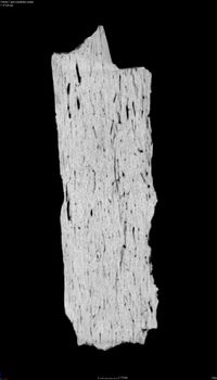
The collection comprises the ZooMS dataset, 3 OBJ files for use with 3D viewer software, and 774 CT scan images in JPEG format that contain 3 separate slices or views (Front, Right and Top).

The CT scan was undertaken using a Nikon XT H 225 micro-scanner with a transmission target. Attempts to keep the dosage as low as possible were made in order to avoid any damage to the sample, so the scan was run at 70kv and 80μA. In total 1,448 projects were taken at two frames per projection, with an exposure set at 100ms and magnification at x7.2. CT data was reconstructed using CT Pro 3D software, and processed with VG Studio Max 2.1 software.


ADS logo
Data Org logo
University of York logo