Facial reconstruction; forensic sciences; 3D modelling; Cyberware; Open Inventor
Contemporary developments in three-dimensional (3D) digitised image capture, graphical modelling and animation have begun to impinge on some quite traditional areas of the forensic sciences. Forensic facial reconstruction serves as a case in point, but surveying and reconstruction and modelling and animation of accident or crime scenes are other fields in which 3D computerised methods are gradually being adopted. A virtual reality modelling language (VRML) 3D crime scene model is now available on the World Wide Web (WWW) � to those who have a 'plug-in' that will read it.
In this article, I would like to concentrate on 3D facial reconstruction, briefly describing the traditional method, and outlining the novel approach being adopted in the computerised 3D forensic facial reconstruction project at the University of Sheffield.
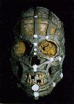
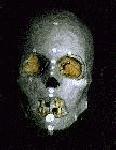
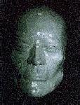
The purpose of forensic facial reconstruction is to produce an image from a skull which offers a sufficient likeness of the living individual that it will facilitate identification of skeletal remains when there are no other means available. Although facial reconstruction had begun in the nineteenth century, the method gained notoriety with the work of Gerasimov (1968), depicted on film in Gorky Park. These traditional 'plastic' methods (Is�an and Helmer 1993, Snow et al. 1970) use modelling clay or plasticine to build up the depth of tissue on the skull (or a cast of the skull) to that of a living individual. Tissue depths are known for 'landmark' sites on the skull; the depths elsewhere are interpolated between these points (Figure 1) and then into the interstices (Figure 2). The shape of the eyes, nose and mouth cannot be confidently predicted and are largely guesswork (Figure 3). Even for skilled practitioners, plastic reconstructions take one or two days. The results obtained will differ between reconstructions and between practitioners.
 Left to right:
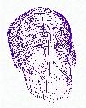
Figure 1 Establishment of tissue depths at landmark sites on the skull (in white) and the interpolation between these sites.
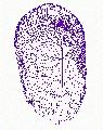
Figure 2. Interpolation of tissue depths into the interstices.
Figure 3. Completed "plastic" reconstruction. The shape of the eyes, nose and mouth are guesswork. [Click on figures to enlarge to full size.]
Left to right:
Figure 1 Establishment of tissue depths at landmark sites on the skull (in white) and the interpolation between these sites.
Figure 2. Interpolation of tissue depths into the interstices.
Figure 3. Completed "plastic" reconstruction. The shape of the eyes, nose and mouth are guesswork. [Click on figures to enlarge to full size.]
The tissue depth measurements used tend to be those collected from cadavers in the early part of the twentieth century, or before. These measurements are biased because they come from small samples, because a dead person's tissues are not the same as in life, and because they take only limited account of the average differences known to occur between people of different age, build and sex, and between the major human diversity aggregates. For over a century, forensic artists and scientists have been attempting to improve the quality of facial reconstructions from the skull, efforts which have met with very limited success. Most recently, computerised methods for 3D facial reconstruction have been developed (Ubelaker and O'Donnell 1992, Vanezis et al. 1989, Shahrom et al. 1996, Miyasaka et al. 1995). These methods employ computer programs to transform laser-scanned 3D skull images into faces. Although the results are more reproducible than sculpted reconstructions, some subjectivity can remain in the 'pegging' of a composite facial image onto the digitised skull matrix. The use of such a standardised image will reduce the influence of the individual shape of each skull, which is after all fundamental to the person's appearance. Computerised methods may be repeatable, fast and precise, but as long as they employ the old data, the quality of the reconstruction will be undermined.
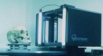 Figure 4. The Sheffield facial reconstruction suite: laser scanner, PC server and Indy� graphics workstation. [Click to enlarge to full size.]
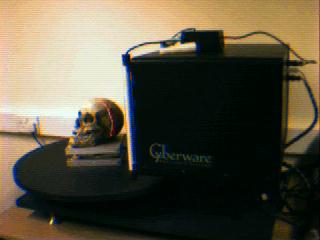 Figure 5. The laser beam directed onto the skull during scanning.
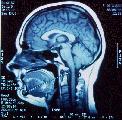
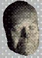
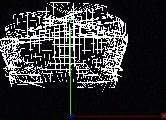
We use a Cyberware 3030 RGB CN color laser scanner and Silicon Graphics Indy� computer to capture 3D images of the skull (Figure 4). As the platform rotates (Figure 5) a 'wireframe' matrix is generated (Figure 6). Computed tomography (CT) scanning permits more accurate measurement of tissue depths (Figure 7). Large samples of tissue depth measurements can be collected, with associated attributes of age, sex, build and, where appropriate, ethnic group. A pilot study on the collection of tissue depth measurements from CT scans has been carried out by one of our team (Nelson 1996). Digitised images of facial features not predicted by the skull contours (nose, eyes and mouth) must be added by separate means to generate a wireframe face (Figure 8), onto which colour and texture can subsequently be rendered (Figure 9). If necessary, a skull can be reconstructed 'virtually' from the separately scanned parts (Figure 10).
We are using C++ and Silicon Graphics' Open Inventor� (Wernecke 1994) programming languages to provide a coherent Windows-based medium for scanning, facial reconstruction and display. We are building features into our model for the selective application of tissue depth datasets defined by parameters such as age, sex, build or ethnic origin. Another component will allow the selection of facial features from computerised image libraries. The graphic and animation facilities of Open Inventor� will allow the display of a moving facial image cycling through a range of versions in a variety of lighting conditions � with the aim of maximising the likelihood of recognition. The animated 3D reconstruction could be downloaded to videotape or, as the Open Inventor� .iv file format is the basis of VRML, the reconstruction could be made available on the Internet to anyone with suitable viewing software � and the password!
Currently, data collection from CT scans is a laborious and time-consuming process of amassing a series of point-to-point measurements. The automated capture of 3D data from CT scans is an imminent proposition, but will not be cost effective for facial reconstruction in the near future. Methods for ageing a reconstruction or for making comparisons with digitised missing persons databases are also far off. It has been suggested that an eventual understanding of the developmental genetics of facial growth may allow forensic or ancient DNA to be used to inform reconstructions. This imaginative idea somewhat downplays the importance of epigenetic factors in physiological development � and the influence of ageing, build and a plethora of environmental influences upon one's appearance! Research on the psychology of face recognition will be applicable, however (e.g. Bruce 1991), and portable laser scanning equipment is shortly to be made available � raising the possibility that computerised facial reconstruction could be used in sensitive human rights work in the field.
Historical facial reconstructions have a precedent going back at least as far as their forensic counterparts. In the nineteenth century, reconstructions were made of the writer-philosopher Schiller and the philosopher Kant (Welcker 1883). More recently, Richard Neave reconstructed the face of a skull from Vergina, reputed to be that of Philip II of Macedonia (Neave 1984), and of Lindow Man (Neave 1989). Neave recently appeared on television depicting a 'Mayan' face belonging to one of the celebrated crystal skulls. However, bearing in mind that the shape of the nose can be predicted with only about sixty per cent accuracy and the shape of the tip with only about forty per cent (Macho 1989, 1986), and that the shape of the hair and hairline is unknown, this was indeed an inspired reconstruction!
Many of Neave's reconstructions are exhibited at the Manchester Museum. Exhibitions of reconstructions have also proved popular at Jorvik in York (where facial imaging techniques are used) and at the Museum nan Eilean, Stornoway (MacLeod and Cowie 1996).
Facial reconstruction is destined to remain an art, albeit an increasingly informed one. The shape of the face bears only a restricted resemblance to the underlying bone structure. Facial reconstructions are inherently inaccurate, therefore, and cannot be used as a positive proof of identification � certainly not in a court of law. Like many things in archaeology, a facial reconstruction is a scientifically-informed artistic recreation � an interpretation. Nevertheless, a forensic facial reconstruction has value in potentially allowing the exclusion of a particular individual as the unidentified subject and, most importantly, in acting as a stimulus for recollection of an absent neighbour, friend or relative. In this sense, the accuracy of the image may not be as important as allowing the investigating agencies to benefit from timely media attention and the public eye.
The contemporary relevance of research on computerised modelling and animation may benefit archaeology intellectually and financially. Within archaeology, the enhanced cosmetic aspects of facial reconstruction further increase the amount of error incorporated into the model. Nevertheless, the drama of sensitively presented facial reconstructions may foster a greater empathy with the people whose lives we hope somehow to represent.
Archaeology continuously faces choices about how to engage with the contemporary world. In this, the first issue of Assemblage, I would like to advocate a strategy of a critical appropriation of science and technology for archaeology.
The Sheffield computerised 3D forensic facial reconstruction project team includes Andrew Chamberlain, Beth Rega, Andy Tyrrell, Martin Evison, Linda Nelson and Kate Howell, and is directed by Prof. Michael Green of the Department of Forensic Pathology. We thank Phil Robinson of St. James' Hospital, Leeds for access to CT facilities. I thank Ian Tyers of Sheffield Dendrochronology Laboratory for his tireless scanning. The project is supported by the UK Home Office.
Bruce, V. 1991. ed. Face recognition. London: Erlbaum.
Gerasimov, M.M. 1968. The Face Finder. London: Hutchinson and Co.
Is�an M.Y. and Helmer, R.P. 1993, eds. Forensic Analysis of the Skull. New York: Wiley Liss.
Macho, G. 1986. An appraisal of plastic reconstruction of the external nose. Journal of Forensic Sciences, 31, 1391-403.
Macho, G. 1989. Descriptive morphological features of the nose � an assessment of their importance for plastic reconstruction. Journal of Forensic Sciences, 34, 1021-36.
MacLeod, I. and Cowie, T. 1996. A face from the past. Current Archaeology, 147, 100-1.
Miyasaka, S., Yoshino, M., Imaizumi, K. and Seta, S. 1995. The computer-aided facial reconstruction system. Forensic Science International, 74(1-2), 155-65.
Neave, R.A.H. 1984. The skull from tomb II at Vergina: King Philip II of Macedon. Journal of Hellenic Studies., 104, 60-78.
Neave, R.A.H. 1989. Reconstruction of the skull and the soft tissues of the head and face of Lindow Man. Canadian Society of Forensic Science Journal, 22, 43-53.
Nelson, L.A. 1995. The Potential Use of Computed Tomography Scans for the Collection of Cranial Soft Tissue Depth Data [M.Sc. dissertation, unpublished]. Sheffield: University of Sheffield.
Shahrom, A.W., Vanezis, P., Chapman, R.C., Gonzales, A., Blenkinsop, C. and Rossi, M.I. 1996. Techniques in facial identification: computer-aided facial reconstruction using a laser scanner and video superimposition. International Journal of Legal Medicine, 108(4), 194-200.
Snow, C.C., Gatliff, B., and McWilliams, K.R. 1970. Reconstruction of facial features from the skull; an evaluation of its usefulness in forensic anthropology. American Journal of Physical Anthropology, 33, 221-8.
Ubelaker, D.H. and O'Donnell, G. 1992. Computer assisted facial reconstruction. Journal of Forensic Sciences, 37, 155-62.
Vanezis, P., Blowes, R.W., Linney, A.D., Tan, A.C., Richards, R. and Neave, R. 1989. Application of 3-D computer graphics for facial reconstruction and comparison with sculpting techniques. Forensic Science International, 42, 69-84.
Welcker, H. 1883. Schiller's Sch�del und todenmaske, nebst mittheilungen �ber Sch�del und todenmaske Kants. Braunschweig.
Wernecke, J. 1994. The Inventor mentor: programming Object-oriented 3D graphics with Open Inventor� , release 2. New York: Addison-Wesley.
�Martin Evison 1996

�assemblage 1996